Tnm Mesothelioma Radiology - The 4 Mesothelioma Stages Explained / This new staging system, in combination with additional tumor factors such as histologic type and patient demographics (eg, age at diagnosis and performance status), is used to determine prognosis.
Tnm Mesothelioma Radiology - The 4 Mesothelioma Stages Explained / This new staging system, in combination with additional tumor factors such as histologic type and patient demographics (eg, age at diagnosis and performance status), is used to determine prognosis.. The primary differential considerations include pleural metastases, pleural dissemination of thymoma, localized fibrous tumor of the pleura, and epithelioid hemangioendothelioma. Three histologic types of mpm are seen at microscopy (13). Up to 25% of patients have metastatic disease at the time of presentation if staged with fdg pet 5. Additional risk factors for mpm include radiation therapy for other malignancies (eg, breast and lung cancers) and simian virus 40, an oncogenic deoxyribonucleic acid virus whose role in the development of mpm is controversial (7). ■ identify specific risk factors for mpm.
Additional risk factors for mpm include radiation therapy for other malignancies (eg, breast and lung cancers) and simian virus 40, an oncogenic deoxyribonucleic acid virus whose role in the development of mpm is controversial (7). Mpm may rarely demonstrate osseous and/or cartilaginous differentiation (15). See full list on radiopaedia.org The abbreviation tnm signifies three different parts of a mesothelioma diagnosis. David sugarbaker developed the brigham staging system, which focused on surgery and whether a patient is eligible at different stages of progression.
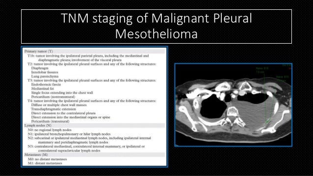
Malignant Pleural Mesothelioma Pulmonology Advisor from www.pulmonologyadvisor.com Not all types of asbestos are strongly implicated, with crocidolite being the main causative fiber type. See full list on radiopaedia.org Pleural opacity which may extend around and encase the lung 2. See full list on pubs.rsna.org ■ correlate imaging findings with the staging of mpm and understand the implications for therapy and survival. Therefore, it is important for the radiologist to understand the strengths and limitations of ct, mr imaging, and pet/ct in the evaluation and staging of patients with mpm. We review the tnm staging system because it emphasizes factors related to overall survival rate, including the local extent of primary tumor (t), lymph node involvement (n), and metastatic disease (m) (tables 1, 2). The most common imaging manifestations of mpm include pleural effusion, pleural thickening, ipsilateral volume loss, local invasion, lymphadenopathy, and metastatic disease.
Although a combination of key imaging features is typically sufficient to suggest a diagnosis of mpm, especially in the appropriate clinical setting, an awareness of other disease processes that may manifest similarly at radiologic examination is necessary.
David sugarbaker developed the brigham staging system, which focused on surgery and whether a patient is eligible at different stages of progression. See full list on radiopaedia.org Over time, mesothelioma staging systems will continue to offer more defined diagnoses, allowing physicians to treat patients with more significant effect and higher chances of survival. See full list on radiopaedia.org A modified version of this system loosely defines the stages of peritoneal mesothelioma as well. Up to 25% of patients have metastatic disease at the time of presentation if staged with fdg pet 5. As researchers gather more knowledge on the progression of malignant pleural mesothelioma, the criteria that define each stage may change again. There is a strong association with exposure to asbestos fibers (~10% risk during lifetime; Three histologic types of mpm are seen at microscopy (13). See full list on pubs.rsna.org Doctors use staging systems to track and measure the progression of cancer. ■ describe the radiologic appearance of mpm across multiple imaging modalities. See full list on asbestos.com
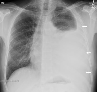
See full list on radiopaedia.org The most common symptoms reported at presentation by patients with mpm are nonpleuritic chest wall pain and dyspnea. A unilateral pleural effusion is seen in up to 74% of patients (fig 1b). Below is the eighth edition of the tnm staging system for malignant pleural mesothelioma , which was published in 2018 1. However, improved survival and decreased morbidity and mortality have been demonstrated when the diagnosis is made in the early stages of disease and when specific treatment strategies are used.
02 Pleural Disease 2019 Radiology from image.slidesharecdn.com David sugarbaker developed the brigham staging system, which focused on surgery and whether a patient is eligible at different stages of progression. The most common imaging manifestations of mpm include pleural effusion, pleural thickening, ipsilateral volume loss, local invasion, lymphadenopathy, and metastatic disease. An evolving staging system equips doctors with the knowledge to offer treatment plans tailored to distinct stages. Although a combination of key imaging features is typically sufficient to suggest a diagnosis of mpm, especially in the appropriate clinical setting, an awareness of other disease processes that may manifest similarly at radiologic examination is necessary. Although individual imaging findings may be nonspecific for mpm, the presence of one or more of these features should raise suspicion for mpm, especially in the appropriate clinical scenario. Tumor like conditions of the pleura 6.1. Some areas of the world have very regional hotspots, such as belfast in northern ireland, due to the historic shipbuilding industry. Mpm may rarely demonstrate osseous and/or cartilaginous differentiation (15).
How is the tnm system used in pleural mesothelioma?
See full list on radiopaedia.org We review the tnm staging system because it emphasizes factors related to overall survival rate, including the local extent of primary tumor (t), lymph node involvement (n), and metastatic disease (m) (tables 1, 2). ■ identify specific risk factors for mpm. Stages 1 and 2 are early in the disease process, and stages 3 and 4 involve widespread tissue involvement or multiple tumors. Over time, mesothelioma staging systems will continue to offer more defined diagnoses, allowing physicians to treat patients with more significant effect and higher chances of survival. As researchers gather more knowledge on the progression of malignant pleural mesothelioma, the criteria that define each stage may change again. See full list on radiopaedia.org See full list on pubs.rsna.org Tumor like conditions of the pleura 6.1. Reduced volume of the affected hemithorax, resulting in ipsilateral shift of the mediastinum (common) 4 3. An evolving staging system equips doctors with the knowledge to offer treatment plans tailored to distinct stages. In a postmortem study of 318 patients, 55% patients were found to have extrathoracic metastases, the commonest sites being the liver (32%), spleen (11%), thyroid (7%) and brain (3%) 16. Most commonly is unilateral and exudative or hemorrhagic in nature, with frozen hemithorax (not causing mediastinal shift) ct is most commonly used for imaging assessment of mesothelioma, and sufficient for accurate staging of disease in most patients.
In a postmortem study of 318 patients, 55% patients were found to have extrathoracic metastases, the commonest sites being the liver (32%), spleen (11%), thyroid (7%) and brain (3%) 16. Pleural fibrosisfrom infective/inflammatory source (e.g. A unilateral pleural effusion is seen in up to 74% of patients (fig 1b). Although a combination of key imaging features is typically sufficient to suggest a diagnosis of mpm, especially in the appropriate clinical setting, an awareness of other disease processes that may manifest similarly at radiologic examination is necessary. This new staging system, in combination with additional tumor factors such as histologic type and patient demographics (eg, age at diagnosis and performance status), is used to determine prognosis.
Malignant Mesothelioma Imaging from img.medscapestatic.com Over time, mesothelioma staging systems will continue to offer more defined diagnoses, allowing physicians to treat patients with more significant effect and higher chances of survival. Pleural mass or nodular thickening of soft tissue attenuation 1.1. See full list on radiopaedia.org Other pleural based tumors 6. Tends to cause inward contraction of the hemithorax, e.g. See full list on radiopaedia.org Malignant pleural mesothelioma (mpm) is the most common primary malignancy of the pleura and the second most common overall malignancy of the pleura after metastatic disease (1). See full list on asbestos.com
It is also called the imig staging system after the international mesothelioma interest group, which is the organization that developed it.
Although individual imaging findings may be nonspecific for mpm, the presence of one or more of these features should raise suspicion for mpm, especially in the appropriate clinical scenario. The tumor may spread along the interlobar fissures (fig 2a). An evolving staging system equips doctors with the knowledge to offer treatment plans tailored to distinct stages. See full list on radiopaedia.org Rib destruction or extension beyond the lateral and anterior margins of the chest wall 4. Mpm arises from the mesothelial cells that cover the lung and chest wall and is strongly associated with asbestos exposure, with latency periods ranging from 20 to 50 years. Three histologic types of mpm are seen at microscopy (13). Staging systems translate these results into a number system that oncologists use to give cancer a "stage." there are four stages of pleural mesothelioma: How are chest radiographs used to diagnose mesothelioma? Below is the eighth edition of the tnm staging system for malignant pleural mesothelioma , which was published in 2018 1. David sugarbaker developed the brigham staging system, which focused on surgery and whether a patient is eligible at different stages of progression. See full list on radiopaedia.org Chest ct alone is often sufficient for disease staging and treatment planning.


0 Comments